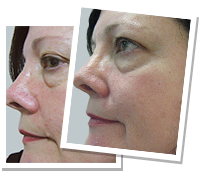
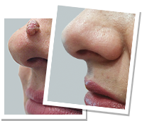
Mole removal before and after
People considering mole removal usually want to see one thing: what the site looks like afterwards. That is a reasonable question, and this page answers it as honestly as a page can.
Photographs are shown in consultation
Before and after photographs of our own patients are shown at your consultation, where cases can be chosen to match your lesion, its site, your skin type and the method proposed - and where we can talk through what did and did not change.
A website gallery is selected to impress. A case shown in consultation can be selected to inform, including one where the scar is more visible than the person hoped.
What the rules require of any before and after image
Under AHPRA's advertising guidelines for regulated health services, before and after images must be of actual patients of the practice, taken under consistent conditions (same lighting, angle, distance, pose), unaltered beyond cropping, labelled with the time elapsed, accompanied by a statement that results vary, and used with the patient's documented consent. They must not create an unreasonable expectation of benefit, and they must not be used as testimonials.
Images marked "not our patients" illustrate a technique. They are not evidence of what a clinic achieves.
What the site actually looks like, by method
| Method | Immediately | At 2-4 weeks | Long term |
|---|---|---|---|
| Radiofrequency (electrosurgery) | Small raw area, may look hot or sunburnt | Scab separated, flat pink patch | Flat mark, usually lightening toward normal tone over months |
| Laser | Darkening or crusting of the lesion | Crust gone, pink or pale patch | Flat mark; some pigment change can persist |
| Cryotherapy | Blister, then scab | Scab separated, pink patch | Higher chance of a permanent pale mark |
| Shave excision | Shallow wound, dressing | Healed, flat pink patch | Flat mark, sometimes slightly lighter than surrounding skin |
| Ellipse excision | Sutured line, longer than the mole itself | Sutures out, raised pink line | A permanent line scar, fading over 12-18 months |
That last row matters. Surgical excision trades a mole for a line, and the line is always longer than the mole was - that is how the wound closes without a bunched edge. On the face the line is planned to follow a natural crease. It fades; it does not vanish.
The comparison that is not on this page
You will not find images here headed "scarless removal", because there is no such thing. Every method of removing a mole leaves some mark. Non-incisional methods usually leave a flat patch rather than a line, which is often less noticeable - but it is still a change to the skin, and it can be lighter or darker than the skin around it.
Anyone showing you a mole removal result described as leaving no trace is either photographing at a flattering angle or making a claim that cannot be supported. See mole removal and scarring.
How to read the images you are shown
- How long after? A site at six weeks looks very different from the same site at twelve months. Scars are at their most obvious around six to eight weeks and improve for a year or more.
- Same lighting and angle? Raking light exaggerates a scar; flat frontal light hides one.
- Same site, same skin type? A result on a shoulder tells you little about a result on a cheek, and vice versa.
- Same method? Excision results and laser results are not comparable.
- Was anything else done? Ask.
What influences your result
- The method used - which is a clinical decision based on what the lesion is, not a preference
- Site: the face generally heals well; the chest, shoulders and upper back scar more heavily
- Size and depth of the lesion
- Your skin type and tendency to keloid or hypertrophic scarring
- Sun exposure during healing - the biggest thing under your control
- Whether you pick the scab
- Infection or wound separation
- Age, smoking and general health
The thing that matters more than the photograph
Choosing a method by which leaves the least mark is the wrong starting point if the lesion has not been assessed. Laser, cryotherapy and radiofrequency destroy the tissue. If a lesion turns out to have been an early melanoma, there is nothing left to send to pathology and no record that it existed.
Anything with features of concern is excised whole and tested - accepting a line scar in exchange for a diagnosis. See skin cancer and mole removal.
Risks
- Scarring - permanent with excision, possible with any method
- Changes in pigmentation at the site, which may persist
- Infection
- Bleeding
- Delayed healing or wound separation
- Keloid or hypertrophic scarring in those prone to it
- Numbness or altered sensation
- Incomplete removal and recurrence
- Missed diagnosis where a lesion is destroyed rather than examined
Booking
Me Clinic, 4 Burke Road, Malvern East VIC 3145. Phone 1300 852 050 or (03) 9569 3511, or use the online consultation form. As at September 2024 and including GST, a mole removal consultation with a Cosmetic Doctor is $150.
Related: mole removal, mole removal guide, scarring, cost.
Individual results may vary. Any surgical or invasive procedure carries risks. Before proceeding you should seek a second opinion from an appropriately qualified health practitioner.
Images on This Page
-
https://www.meclinic.com.au/wp-content/uploads/2024/03/meclinic-logo.png
Me Clinic
-
https://www.meclinic.com.au/wp-content/uploads/2022/02/call.png
call
-
https://www.meclinic.com.au/wp-content/uploads/2022/03/cs4-Before_Post_MOLE-200x171_AB.png
(no alt text)
-
https://www.meclinic.com.au/wp-content/uploads/2022/03/cs5-Before_Post_MOLE-200x171_VK.png
(no alt text)
-
https://www.meclinic.com.au/wp-content/uploads/2024/02/acps.png
(no alt text)
-
https://www.meclinic.com.au/wp-content/uploads/2024/02/racs.png
(no alt text)
-
https://www.meclinic.com.au/wp-content/uploads/2024/02/fracs.png
(no alt text)
-
https://www.meclinic.com.au/wp-content/uploads/2024/02/isaps.png
(no alt text)
-
https://www.meclinic.com.au/wp-content/uploads/2024/02/cpca.png
(no alt text)
-
https://www.meclinic.com.au/wp-content/uploads/2022/02/auscop.png
(no alt text)
-
https://www.meclinic.com.au/wp-content/uploads/2022/02/amav.png
(no alt text)
-
https://www.meclinic.com.au/wp-content/uploads/2022/03/channel7.png
(no alt text)
-
https://www.meclinic.com.au/wp-content/uploads/2022/03/channel9.png
(no alt text)
-
https://www.meclinic.com.au/wp-content/uploads/2022/03/channel10.png
(no alt text)
-
https://www.meclinic.com.au/wp-content/uploads/2022/03/abc.png
(no alt text)
-
https://www.meclinic.com.au/wp-content/uploads/2022/02/malvern-map-thumb.jpg
(no alt text)
-
https://www.meclinic.com.au/wp-content/uploads/2022/03/home-conctact-tab.jpg
(no alt text)
-
https://bat.bing.com/action/0?ti=56339805&Ver=2&mid=76a3db88-aca8-474f-ab27-aca174784d02&bo=1&sid=80859170ac3311f1bde3fded96ffbf7a&vid=8085ae70ac3311f19d71e35c04e9f8a7&vids=0&msclkid=N&pi=918639831&lg=en-US&sw=1280&sh=720&sc=24&tl=Mole%20Removal%20Procedures%20-%20What%20to%20Expect%20and%20Process&p=https%3A%2F%2Fwww.meclinic.com.au%2Fskin%2Fmole-removal%2Fbefore-and-after%2F&r=<=2032&evt=pageLoad&sv=2&cdb=AQAQ&rn=381132
(no alt text)